Vitamin D Toxicosis in Pets
The ASPCA’s Animal Poison Control Center reports that more potent supplements are becoming widely available to address an increased concern for Vitamin D deficiency in humans, so veterinarians are encountering more Vitamin D toxicosis in companion animals.
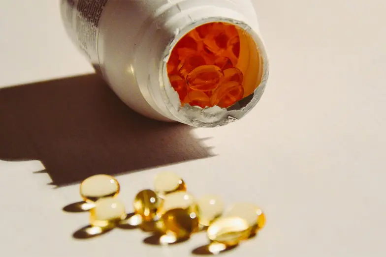
Cholecalciferol (Vitamin D) toxicosis can come from a variety of different sources, with the most common being supplements. Other sources of Vitamin D ingestions include:
-
Cholecalciferol rodenticides, which are very potent and can cause toxicity with just a small exposure.
-
Psoriasis creams that contain a vitamin D analog, such as calcipotriene. These are highly potent and a very small exposure can cause severe signs of toxicosis.
-
Some plants such as Cestrum diurnum, Trisetum flavescens and Solanum malacoxylon can contain very high levels of Vitamin D. The biggest concern for toxicosis in animals is for herbivores who graze on pastures with these noxious weeds, but any animal can become intoxicated with a sufficient ingestion.
Stay on top of FDA pet food recalls and share the information with your clients.
Cholecalciferol Toxicity & Treatment
Cholecalciferol is metabolized in the liver to 25-hydroxycholecalciferol (calcifediol), which in turn is metabolized in the kidney to 1,25-dihydroxycholecalciferol (calcitriol). Calcitriol is the active metabolite and increases intestinal absorption of calcium, stimulates bone resorption of calcium and increases renal tubular reabsorption of calcium.
This leads to hypercalcemia and hyperphosphatemia.
When the calcium phosphorus product exceeds 60, mineralization of soft tissues can be seen, leading to acute kidney injury. In addition, a ruptured aorta can be seen if the aorta is calcified.
Patients may develop vomiting, diarrhea, inappetance, polydipsia and polyuria, and the signs may be delayed.
Emesis may be recommended for healthy, asymptomatic patients with recent exposures. Activated charcoal can be helpful with adsorbing cholecalciferol. Cholestyramine powder should be considered, as it has been also shown to be effective in lab animals and humans to enhance excretion of vitamin D by binding bile acids.
Baseline calcium, phosphorus, BUN and creatinine should be obtained and monitored for at least four days.
Treatment can be complex and depends on monitoring clinical signs and laboratory values very closely. If changes in bloodwork consistent with exposure occur, treatment may be indicated including:
- 0.9% NaCl fluid diuresis
- Furosemide. Loop diuretics can increase renal clearance of calcium; however, avoid thiazide diuretics as they reduce renal excretion of calcium
- Prednisone. Steroids reduce bone resorption, decrease intestinal absorption and increase renal excretion of calcium
- Magnesium hydroxide is a phosphate binder and can be added to help reduce hyperphosphatemia
- Pamidronate, a bisphosphonate used in humans to treat hypercalemia of malignancy
- Low calcium diet
Patients requiring treatment for hypercalcemia and hyperphosphatemia should be slowly weaned off any treatments. Bloodwork should be monitored until the patient's bloodwork returns to normal and remains normal without any treatments.
GI upset, inappetance, lethargy and PU/PD are indications that bloodwork should be checked, as serum calcium levels may be increasing.
We have lots more on this subject: